Tesera SC Stand-Alone Cervical Interbody System
Tesera SC Stand-Alone Cervical Interbody Fusion System with Tesera Trabecular Technology
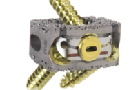
The Tesera® SC Stand-Alone Cervical Interbody Fusion System combines our most advanced material science and revolutionary manufacturing process into a powerful stand-alone cervical interbody fusion system.
Using additive manufacturing, we have created an ideal trabecular structure for bone in-growth, providing excellent long-term fixation. Three screws and an innovative locking system provide immediate fixation and confidence.
Tesera SC Stand-Alone Cervical Interbody Fusion System with Tesera Trabecular Technology
KYOCERA Medical is committed to advancing patient care through better products, based on scientific and clinical data. We commissioned an independent study through IMDS Discovery Research to validate our Tesera technology. Below are images and excerpts from the Twelve Week Draft Report. The final report, with 24 Week data, will be posted upon completion.
A Twelve and Twenty-four Week Study to Quantify the Rate of Bone Growth into a Porous Titanium Device in the Ovine Medial Femoral Condyle.
By: IMDS Discovery Research | 1785 North 730 West | Logan, Utah 84321
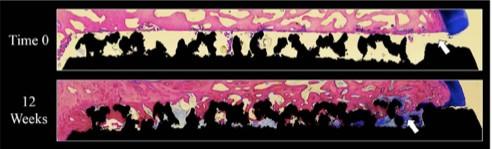
Figure 12: This figure shows an overhead view of the 75 micron stained sections. Black=Titanium, Pink=Bone, Blue=Fibrous Tissue & White=Pore Space. The 12 Week specimen had excellent bone attachment to the porous coating. Note – no bone grafts, synthetics or other extenders were used in this study.
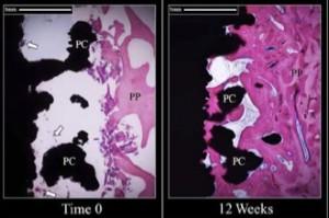
Figure 13: This figure shows a comparison of each time point. Black=Titanium, Pink=Bone, Blue=Fibrous Tissue & White=Pore Space. The time 0 specimen shows bone chips (arrows) within the porous structure (PC) and along the drill track in the periprosthetic region (PP). 12 Week specimen shows excellent bone attachment to the porous structure.
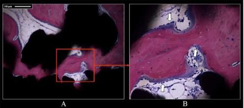
Figure 14: This figure shows bone ingrowth and remodeling within the porous structure on the 12 week specimen. Black=Titanium, Pink=Bone, Blue=Fibrous Tissue & White=Pore Space. (A) 10x image showing bone attachment to the porous structure, (B) Higher power image of (A) (red box) showing osteoblastic activity (arrows) within the porous coating.
Features and Benefits
The Tesera SC – Stand-alone Cervical Interbody Fusion System combines our most advanced material science and revolutionary manufacturing process into a powerful stand-alone cervical interbody fusion system.
01 Cages
- Tesera Trabecular Technology – allows bone on-growth and ingrowth
- Triple screw fixation for immediate stability
- Single-step locking plate secures all 3 screws with tactile and visual locking confirmation
- Parallel and 7˚ Lordotic
- Heights from 6mm – 12mm
- Two footprints
02 Instruments
- All-in-one Drill Guide for one-step insertion and fixation
- Straight and U-jointed instruments for hassle-free screw placement
- Trials and Broaches for every cage size
03 Screws
- Fixed and Variable screws
- Self-drilling and Self-tapping options
- 3.5mm or 4.0mm diameters
- Lengths from 12mm – 18mm (2mm increments)
Sizes and Specs
Tesera SC Stand-alone Cervical Interbody Fusion System has two footprints.
